Origins and structure of the diatom chloroplast. Steinii the chloroplast is generally associated with pyrenoid covered with starch plates but sometimes pyrenoids can be more than one.

Ultrastructure Of Aulacoseira Baicalensis Chloroplast A B Lm A Download Scientific Diagram
The mycelium is coenocytic and branched.

Diatom diagram chloroplast. Suggests cross-talk between mitochondria and chloroplasts. Among them diatoms and dinoflagellates are the two most common phytoplankton species that can be found in. Brightfield x Keep in mind that the cutaway diagram of a diatom is a very generalized schematic and cannot adequately describe the variation in all diatoms.
Chloroplast anatomy is the chloroplast structurechloroplast measure 2um 5um in diameter. The chloroplast is H shaped in C. CO 2 and HCO 3 are treated in four compartments extracellular solution cytoplasm chloroplast stroma and pyrenoid subject to hydration and dehydration within each compartment indicated with dark blue arrows photosynthetic removal in the pyrenoid green arrow and fluxes between compartments.
In this article we will discuss about the life cycle of saprolegnia with the help of suitable diagrams. They generate cell movement through cytoplasm that streams along the raphes always. Exploring the application of mathematics and computer science.
Digital measurement of distances and angles. Create a venn diagram to show the similarities and differences between plant cells and animal. Diatoms are responsible for a large fraction of CO2 export to deep seawater a process responsible for low modern-day CO2 concentrations in surface seawater and the atmosphere.
In diatoms in order to regulate the redox balance of the cell. Mitochondria is found in. I Rhizoidal or intramatrical hyphae.
Alternative oxidase chloroplast diatom mitochondria. The girdle lamella and the thylakoids are always found in bundles of. Shown in the right-hand false-colour picture is a subfossil assemblage from a muddy deposit a.
The key difference between diatoms and dinoflagellates is that the diatoms have a cell wall composed of silica while the dinoflagellates have a cell wall composed of cellulose. Whereas photosystem II provides energy for the water splitting and oxygen releasing. Diatoms are among the most important and prolific microscopic sea organisms and serve directly or indirectly as food for many animals.
This schematic figure shows two alternative hypotheses for the origins of the diatom chloroplast 42542. Diatom class Bacillariophyceae any member of the algal class Bacillariophyceae division Chromophyta with about 16000 species found in sediments or attached to solid substances in all the waters of Earth. Mycelium of Saprolegnia Fig.
As the image of Coscinodiscus shows diatom chloroplasts are usually yellowish-brown in color ranging between yellowish-green and dark brown. These are short hyphae which penetrate. However harmful algal bloom as known as red tide is a major problem in environment and fishery industry.
The construction of the cell wall called the frustule consists of two valves that fit into each other like a little pill box. Diagram of the chloroplast pump model with representative C i concentrations. Diatoms are divided into two groups that are distinguished by the shape of the frustule.
Their cell walls are made of silica almost like a glass house. The diatom chloroplast performs effectively all of the essential pathways identified in plant chloroplasts including photosynthesis central carbon and lipid metabolism synthesis of plastidial amino acids eg glutamate glutamine cysteine lysine branched chain and aromatic amino acids chlorophyll and carotenoid synthesis and essential plastid biogenesis pathways associated with expression of the chloroplast. Diatoms are a group of phytoplankton that is widely distributed in the hydrosphere and even in moist soil.
2 Inside the chloroplast there are two different photosystems each a large complex of different proteins and pigments that is optimized to harvest light. Like other photosynthetic organisms diatoms have adapted to these low ambient concentrations by operating a CO2 concentrating mechanism CCM to elevate the concentration of CO2 at the site of fixation. Each one of their valves have openings that are slits along the raphes and their shells are typically elongated parallel to these raphes.
Pennate diatoms are bilaterally symmetric. Mucicola stellate in C. Basic arithmetic and trigonometric operations use of reciprocals fractions roots exponentiation order of operations.
These chloroplasts are bounded by up to five membranes depending on whether the entire diatom endosymbiont is counted as the chloroplast or just the red algal derived chloroplast inside it. Arachne and axile in C. Bicilliata reticulate in C.
Diatoms play a great role in carbon fixation with about 20 of the whole fixation in the world. Reticulata parietal in C. During the vegetative phase it is composed of two kinds of hyphae.
The chloroplast is a major source for generation of both ROS and reductive power to generate and recycle NADPH thioredoxin and GSH Dietz et al 2016. The pyrenoids are two in. They play important roles in global carbon-oxygen cycles and provide valuable products.
Each cell contains one to several brownish chloroplasts. We propose that in diatoms chloroplast E GSH is involved in sensing specific environmental stress cues and in regulating cell fate. The left-hand picture above shows a spread of living diatoms and other algae from a freshwater loch in Scotland.
Phytoplanktons are algae that are single-celled eukaryotic cells. Anatomy of chloroplasts green algae and plants including green stems and unripe fruit harbor chloroplaststhe vital organelles where photosynthesis takes place. Diatoms are delicate unicellular organisms that have a yellow-brown chloroplast that enables them to photosynthesize.
Complete the venn diagram to compare the stroma and a granum in a chloroplast. To the study of lakes including. Diatom chloroplasts are typical secondary plastids surrounded by four membranes.
The diatom endosymbiont has been reduced relatively littleit still retains its original mitochondria and has endoplasmic reticulum ribosomes a nucleus and of course red algal derived chloroplastspractically. The outer envelope is termed the chloroplast endoplasmic reticulum CER and is continuous with the nuclear envelope. The rerouting of electron s during iron- limiting cond itions.
A recent article in Nature Communications focuses on a most important area of diatom physiology the harvesting of light energy to drive photosynthesis. There are many types of phytoplankton. The centric diatoms and the pennate diatoms.
Even though intensive studies have been conducted so far the molecular mechanism behind harmful algal bloom was not fully understood. There are two major diatoms have been sequenced but. Diatom plastids are also characterized by a girdle lamella that runs in parallel with the four membranes that surround them.

Schematic Overview Of Chloroplast And Thylakoid Structure In Diatoms Download Scientific Diagram

Origins And Structure Of The Diatom Chloroplast This Schematic Figure Download Scientific Diagram

A Schematic Diatom Plastid Surrounded By Four Membranes The Download Scientific Diagram

The Primary And Secondary Endosymbiotic Events That Gave Rise To Modern Download Scientific Diagram

Origin Of Chloroplasts By Secondary Endosymbiosis Involving A Red Algal Download Scientific Diagram

Thalassiosira Proshkinae Chloroplast Ultrastructure A B Light Download Scientific Diagram

The Ppc And Cell Structure Plastid Compartmentalization Of The Diatom Download Scientific Diagram
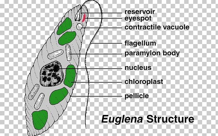
Euglena Cell Protist Diatom Algae Png Clipart Algae Angle Area Cell Chloroplast Free Png Download
Schematic Representation Of The Secondary Endosymbiont Hypothesis Of Download Scientific Diagram
A The Diatom Frustule B A Centric Diatom Showing Several Download Scientific Diagram

Evolution Of Thylakoid Membrane Organization And Composition Download Scientific Diagram
Origins And Structure Of The Diatom Chloroplast This Schematic Figure Download Scientific Diagram

File Euglena Diagram Jpg Wikipedia

Chloroplasts And Mitochondria Are Closely Juxtaposed Inside Diatom Download Scientific Diagram
Diatom Cell Wall Eukaryote Chloroplast Anatomy Transparent Png

Schematic Overview Of Chloroplast And Thylakoid Structure In Diatoms Download Scientific Diagram

Chloroplasts And Mitochondria Are Closely Juxtaposed Inside Diatom Download Scientific Diagram
![]()
The Diatom Ccm Diatom Cells Acquire C Through The Active Transport Of Download Scientific Diagram

Conceptual Schematics Of A A Typical Diatom And B A Smaller More Download Scientific Diagram
إرسال تعليق